‘AI-triaged’ 3D pathology improves cancer detection
An interdisciplinary research team including Wynn Burke, research consultant, and Dr. William Grady, professor (Gastroenterology) recently found a more accurate and less labor-intensive way to diagnose malignancies.
The team began by pioneering a new imaging technology called Open-Top Light Sheet (OTLS) microscopy—in which whole tissue specimens are treated with specific reagents to render them optically clear before being stained and imaged in 3D on a specially-equipped microscope.
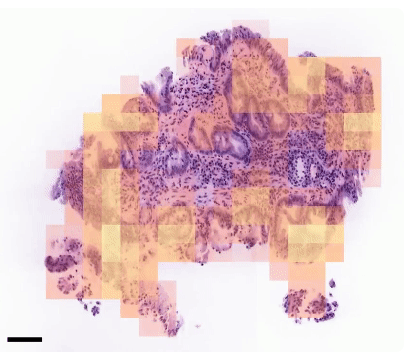
They then sought to streamline the process further, training an AI algorithm to identify different 2D slices of a sample and another algorithm to identify the top three 2D slices (ranked by ‘probability of containing neoplastic tissue’) for pathologist review.
The team calls this ‘AI-triaged’ 3D pathology, and has found it to be successful in improving identification of esophageal neoplasias in patients with Barrett’s esophagus, all while reducing the workload per specimen for the reviewing pathologist.